
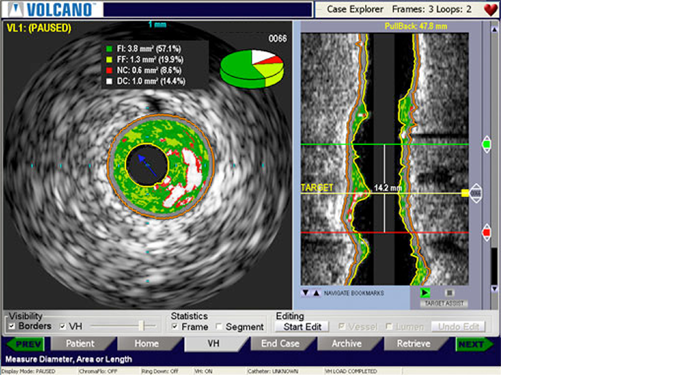
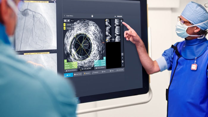
VH IVUS imaging provides a colorized tissue map of plaque composition with automated lumen and vessel measurements.* VH IVUS technology uses advanced, proprietary spectral analysis techniques to classify plaque into four tissue types with 93-97% accuracy.1
Complete lesion assessment
VH IVUS provides a colorized tissue map of plaque composition for complete lesion assessment. Based on more than 15 years of research and 350 scientific publications, including the landmark PROSPECT trial, VH IVUS can assist you with lesion risk assessment, identification of necrotic core for optimizing stent placement, and monitoring of transplant patients for cardiac allograft vasculopathy.
VH IVUS features
Colorized tissue map
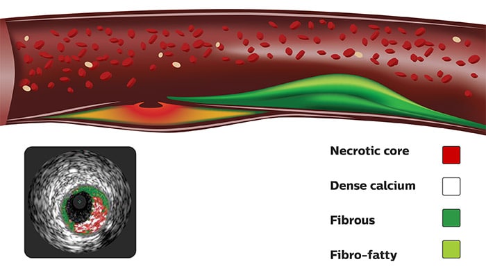
For complete lesion assessment, VH IVUS provides a colorized tissue map of four tissue types:
Necrotic core/dense calcium only mode
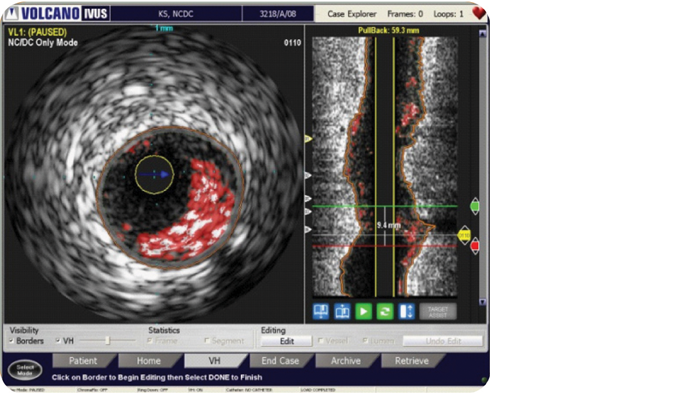
For a simplified view, VH IVUS features an alternate option to display only necrotic core and dense calcium.
Automatic border contours
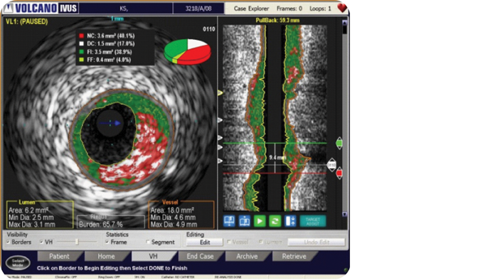
VH IVUS provides automatic border contours for full segment analysis.
Clinical applications for VH IVUS2
Lesion risk and assessment of disease severity2
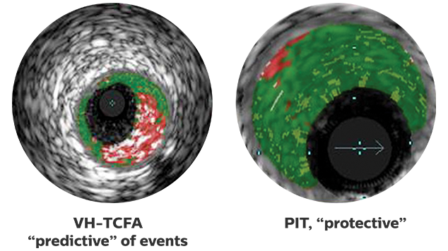
VH IVUS may help assess lesion risk:
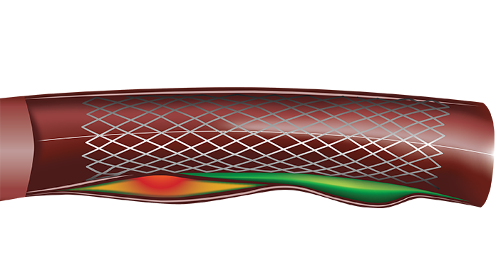
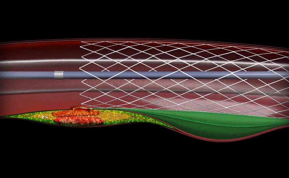
Longitudinal stent placement
Landing stent edges in necrotic core increases the risk of stent thrombosis.3 With VH IVUS, the location of necrotic core can be identified and stent placement optimized.
Culprit of the culprit animation
Related products
-
Eagle Eye Platinum
The Eagle Eye Platinum digital IVUS catheter is the #1 choice of physicians for intravascular imaging (in the US).* As a unique plug-and-play intravascular imaging catheter it is designed for ease of use and deliverability. Features include a soft tapered tip, GlyDx hydrophilic coating for increased lubricity, a long, rapid exchange lumen for improved pushability, three radiopaque markers, and compatibility with SyncVision for co-registration with angiography.
85900P -
Eagle Eye Platinum ST
The Eagle Eye Platinum ST digital IVUS catheter offers a 2.5 mm tip-to-imaging distance designed to assess more of the vessel than standard catheters by providing a closer visualization of highly stenosed lesions and distal anatomy. The short tip catheter fits through all 5F guides and has all features of our top-selling Eagle Eye Platinum model, including plug-and-play simplicity, three radiopaque markers, GlyDx hydrophilic coating, and SyncVision compatibility.*
85900PST -
Core
The Core precision guided therapy system allows you to have iFR, FFR and IVUS modalities on a single platform.¹ Core helps provide clarity in your approach and gives you confidence in your decisions.
IGTDCORE -
Core Mobile
Core Mobile is designed to power the future of precision guided therapy and offers the choice of imaging and physiology on a single mobile platform.¹ Core Mobile also provides clarity in your approach and confidence in your results by offering additional information during the diagnostic phase, assisting in intervention decisions.
IGTDCOREM


Resources
Reimbursement
*Safety and effectiveness of VH IVUS for use in the characterization of vascular lesions and tissue types has not been established