High-powered gradients boost MR research projects at UKM
- Featuring
- August 30 2024
- 5 min read
- Originally published in February 2023
University Hospital Münster (UKM) radiologists and researchers are among the world’s first to experience what the high gradient power of MR 7700 means for clinical research studies. The team is eagerly exploring the 3.0 Tesla MR system’s potential for supporting their ongoing neuroscience research and innovations in prostate, liver and lymph node imaging. For diffusion imaging in particular, they see markedly higher SNR, shorter scan times and reduced distortions.
At-a-glance:
- At University Hospital Münster, Philips MR 7700 is dedicated to research and was chosen for the strength and quality of its XP gradients and numerous other innovations.
- Exceptional diffusion capabilities benefit the hospital’s prostate program.
- Multiparametric MRI of the prostate is used for diagnosis, but also in MRI/ultrasound fusion guidance for targeted prostate biopsies.
- The power the MR 7700 is also harnessed to support interventions, such as laser interstitial thermal therapy (LITT).

Beginning a new era of outstanding scanning
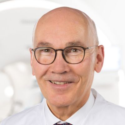
At UKM, the team led by Dr. Walter Heindel operates six MRI scanners. Their latest addition, the Philips MR 7700 is dedicated to research and was chosen for the strength and quality of its XP gradients and numerous other innovations. “We expect that the system will help us to speed up high-end MR imaging even more, without losing image quality,” Dr. Heindel says. “We will concentrate our clinical research projects on this system, especially studies related to patients with neurological and neurosurgical issues and patients with oncologic diseases.” In its first few weeks of operation, more than 100 patients and volunteers have been scanned on the MR 7700. Initial projects for which the team will harness the system include high-resolution multiparametric prostate MRI, developing innovative protocols for stroke imaging, and diffusion studies of the lymph nodes. “We expect that the more powerful gradient system will allow improved diffusion results, meaning more directions and/or more shells [i.e. b-values] and higher SNR without lengthening examination times,” he says. “We also are confident that the Compressed SENSE and MultiBand SENSE acceleration methods will help here. For example, we plan to investigate, whether diffusion techniques in body imaging may serve as an alternative to contrast agent application.”
Improved prostate diffusion images to guide ultrasound biopsies
Dr. Heindel believes the MR 7700 will benefit the hospital’s existing program in which multiparametric MRI of the prostate is not only used for diagnosis, but also for MRI/ultrasound fusion guiding targeted prostate biopsies. “Working with our colleagues in urology, we have been focusing on early detection of prostate cancer,” he says. “Multiparametric MRI helps us identify suspicious lesions. Then fusing the MRI data with ultrasound imaging enables real time MRI-guidance during biopsy, so that urologists can see the lesion and perform a targeted biopsy. We established this interdisciplinary process during the past years to replace conventional blind prostate biopsies. This was also done with Philips and it was a great success.” “From our experience, the diffusion imaging (DWI) seems to provide the most useful information in this process. And it is exactly the diffusion imaging that benefits much from the powerful MR 7700 gradients. So, we will investigate how even higher resolution DWI may enable an innovative approach in this area. The team also aims to investigate multiparametric imaging in combination with machine learning.”
The prostate DWI done so far with the MR 7700 looks promising; the quality of visualizing the gland and the areas of disease seems significantly improved. “In one of our first prostate patients we were already able to acquire a quite high-resolution diffusion image – using a b-value of 3000 – that clearly delineated the prostate lesion. That was a very nice example of what the MR 7700 gradients can achieve,” he says.

Diffusion weighted imaging of prostate. Examples on the left show the regular clinical diffusion protocol with four b-values up to b1000 s/mm². On the right, the T2W image shows a hypointense lesion that has low ADC and is clearly visible in b1400 and b2000 diffusion images, suggesting malignancy.
In one of our first patients we were able to acquire a quite high-resolution diffusion image with a b-value of 3000, which clearly delineated the prostate lesion.

Dr. Walter Heindel
Professor of Radiology and Chair, Director Clinic for Radiology
University Hospital Münster, Germany

High angular diffusion imaging using 257 directions, 23 b-values, maximum b-value 4000 s/mm² in a total scan time of 11 min. These settings allow improved fiber tracking and enhance analysis of intra-voxel incoherent motion (IVIM) and diffusion kurtosis (DK) effects in one single measurement.
Innovating stroke imaging
The UKM is a leading neurovascular center and stroke research is an important activity. DWI’s sensitivity for small and early infarcts of stroke helps in managing these patients.
“Using the MR 7700 we want to develop and test innovative diffusion protocols for stroke imaging to allow fast, confident diagnosis,” Dr. Heindel says. “Apart from critically ill patients, individuals often present here with stroke symptoms for whom we have to quickly assess if they are really having a stroke and need rapid hospitalization in the stroke unit, while in the case of a stroke mimic the patient may be sent home with medication if needed.”
High resolution providing diagnostic confidence
In select cases, MR 7700 has helped the hospital’s physicians more clearly visualize pathology. “We’re definitely getting the impression that tumors are better delineated with the MR 7700,” Dr. Heindel says. “For example, I examined a patient who had been diagnosed in another hospital with possible neuritis of the optic nerve. However, the MR 7700 images allowed me to diagnose it as an optic nerve sheath meningioma, a rare and often misdiagnosed, slowly growing tumor that was causing the visual disturbances in the patient. The lesion was so well delineated on the high-resolution MR 7700 images that our neurosurgeon decided he did not need a biopsy before proceeding directly with decompression of the optic canal and peeling away those tumor cells.”
The images below show how high resolution-MRI impressively demonstrates the compression and narrowing of the right optic nerve in this case of optic nerve sheath meningioma (ONSM). The coronal T2-weighted images show the hyper-intense, half-moon shaped lesion, that is clearly visible in the axial T1W image after contrast injection (right). These imaging findings were so convincing that the responsible neurosurgeon did not consider a pretherapeutic histological clarification.
We’re definitely getting the impression that tumors are better delineated with the MR 7700.
Dr. Walter Heindel
Professor of Radiology and Chair, Director Clinic for Radiology
University Hospital Münster, Münster, Germany

MRI of optic nerve sheath meningioma (ONSM). The coronal T2-weighted images show the hyper-intense, half-moon shaped lesion, that is clearly visible post contrast in the axial T1W (right).
Easy design of imaging protocols and amazing patient comfort
Dr. Heindel has received impressions that MR 7700 operation has been straightforward. “My highly specialized team of MR physicists raves about the ease of use of the system, especially the simple implementation of new imaging protocols. The low effort required to modify scan parameters and protocols enables fast and easy experimentation with imaging techniques,” he says. “This clearly distinguishes Philips and was one of the reasons to choose this system.”
There has also been positive feedback from patients. “One patient told me that she had never had an MRI examination that was so comfortable,” Dr. Heindel says. “By virtue of the softness of the ComfortPlus mattress, combined with the short exam time, she was surprised at how pleasant the experience was, while in earlier examinations at other sites she just wanted to get out of the system. She said she would definitely drive the long distance again to have her follow-up examination also in this system.”

Voxel based morphometry. The neuroscience team compared their standard T1 brain morphometry sequence with alternate protocols facilitating Compressed SENSE. Selected examples are CS shown. All protocols resulted in comparable cortical thickness results with a slight decrease for higher CS-factors.
Expanding research horizons with the MR 7700
University Hospital Münster will also harness the MR 7700 to support interventions, such as laser interstitial thermal therapy (LITT). LITT is a minimally invasive surgical technique to treat brain tumors and epileptic foci by implanting a laser catheter into the tumor or epileptic focus and heating it to temperatures high enough to ablate the disease.
“While depositing heat energy with LITT to destroy pathology, the surgeon has to take care to prevent or limit damage to surrounding normal tissues,” Dr. Heindel says. “We are interested in doing thermometry with the MR 7700 system – measuring body tissue temperatures with MR to find out if the MR 7700 enables faster, more precise monitoring of LITT thermal changes.”
In a neurosurgery project, Dr. Heindel’s team is eager to discover how the MR 7700 gradients might help provide a more comprehensive picture of the brain prior to neurosurgery. “In the preoperative diagnostic work-up, DTI and fMRI are used to find out how close a tumor is to motor cortex areas, and to discover whether fiber tracts go around a tumor or through a tumor,” he says. “We can then transmit these data into the neurosurgeon’s brain imaging suite.” There are also ongoing projects with real-time resting state fMRI.

Finally, in keeping with a worldwide focus on hygienic practices as a result of Covid-19, University Hospital Münster has placed its MR 7700 in a special scan room to allow investigation of fumigation for room cleaning. “This special exam room is equipped with an innovative sterilization technique involving the application of nebulized hydrogen peroxide,” Dr. Heindel says. “This will allow us to investigate whether our approach is feasible to prevent the transmission of pathogens such as bacteria or viruses during radiological examinations. Sterile conditions in hospital environments have never been more important.”
We are interested in doing thermometry with the MR 7700 system to find out if the MR 7700 enables faster, more precise monitoring of LITT thermal changes.
Dr. Walter Heindel
Professor of Radiology and Chair, Director Clinic for Radiology
University Hospital Münster, Münster, Germany
Featuring

Dr. Walter Heindel
MD
Professor of Radiology and Chair, Director Clinic for Radiology
University Hospital Münster, Münster, Germany
Read more
Major fields of research are early detection of disease by imaging (screening, molecular imaging, advanced imaging approaches, population-based research) and minimal-invasive image-guided therapy (interventional oncology and cardio-vascular radiology).
High-powered gradients boost MR research projects at UKM
Copy this URLto share this story with your professional network
Sign up for news and updates

Disclaimer
Results are specific to the institution where they were obtained and may not reflect the results achievable at other institutions. Results in other cases may vary.