Image-guided therapy
Peripheral IVUS
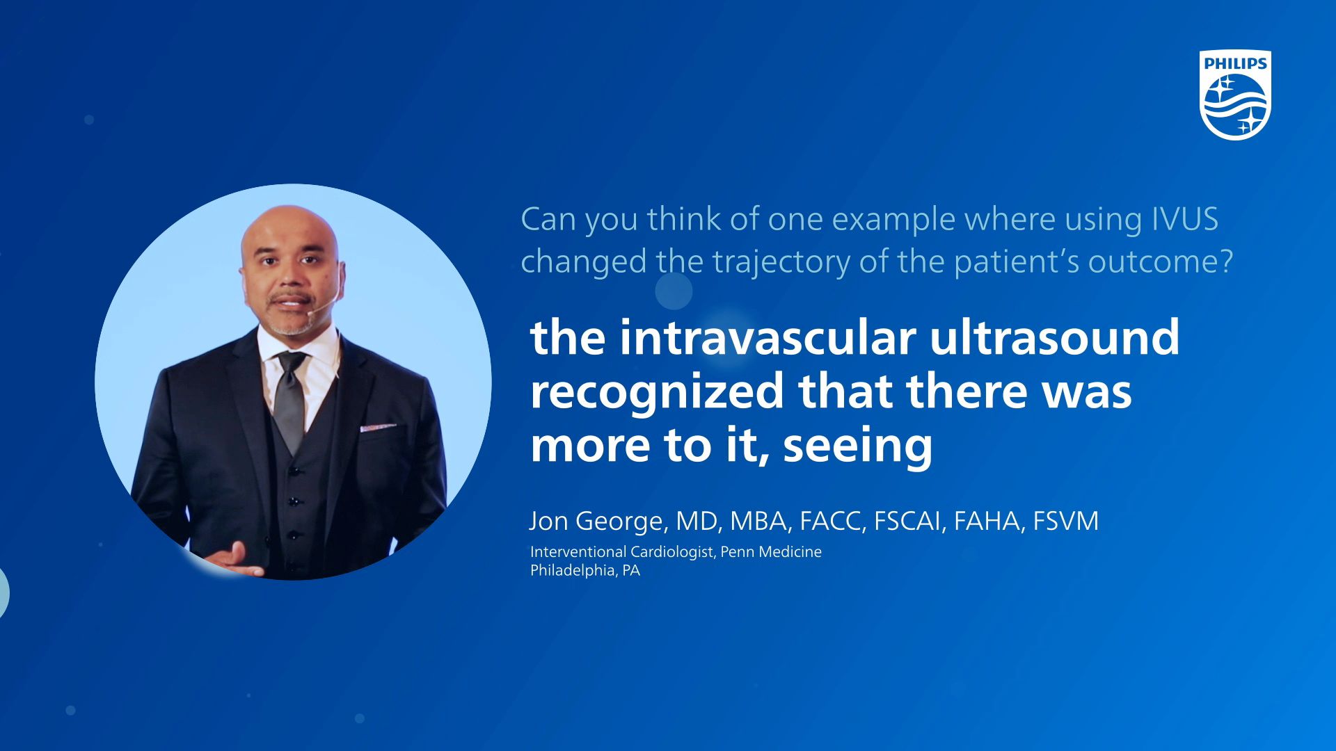
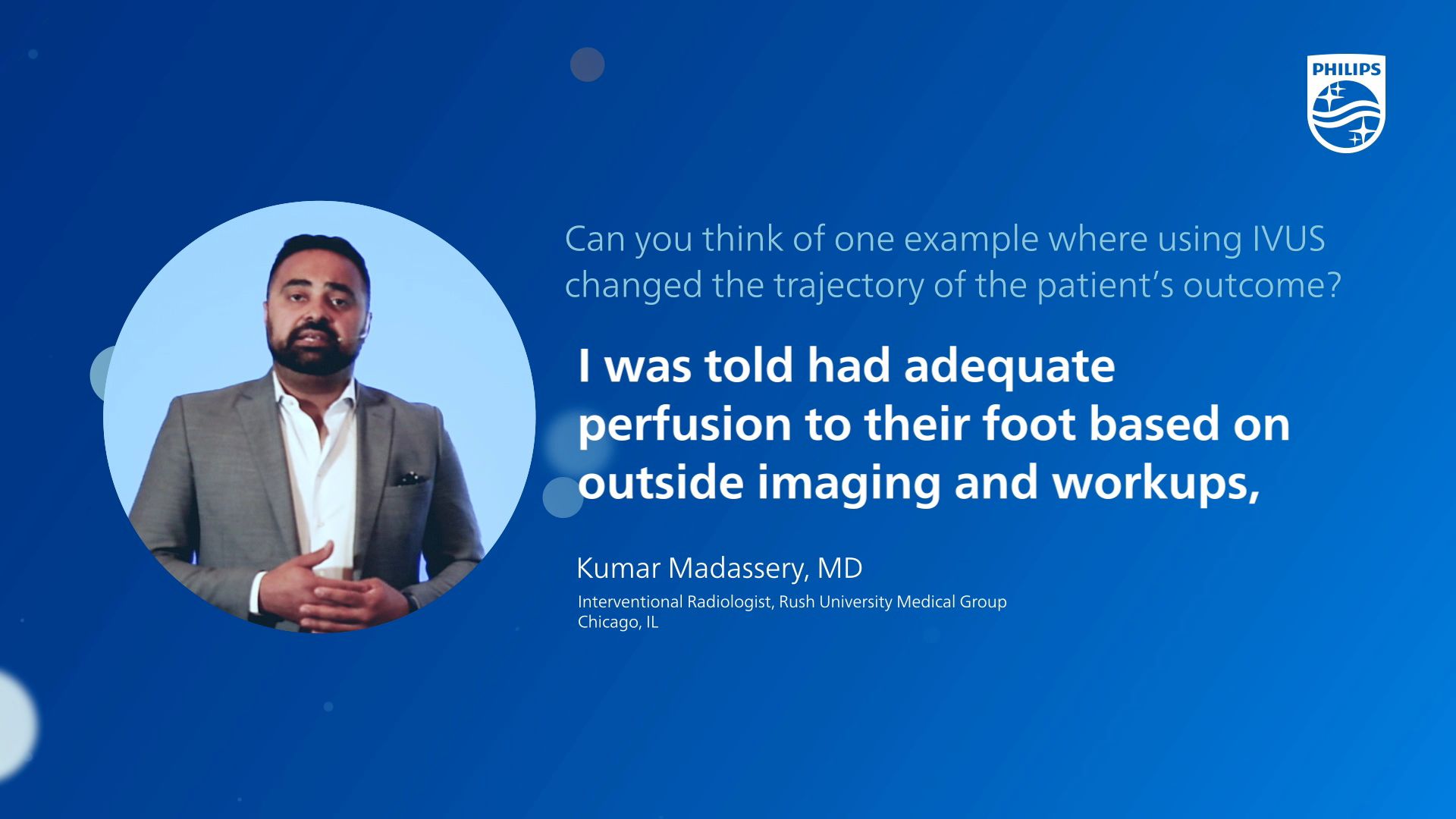
Watch this video to see how IVUS can help you see clearly and treat optimally
27%
reductions in major adverse arterial limb events3
Data from the largest ever real-world clinical analysis of over 700k peripheral arterial and peripheral venous patients on lower extremity peripheral intervention for IVUS, demonstrate a 27% reduction in risk for major adverse limb events including acute limb ischemia or major amputation.
28%
reductions in repeat venous interventions, hospitalization or death4
Data from the largest ever real-world clinical analysis of over 700k peripheral arterial and peripheral venous patients on lower extremity peripheral intervention for IVUS, demonstrate a 28% risk reduction for repeat intervention, hospitalization, or death.
1st
ever global consensus guidance on IVUS5
40 cross-specialty experts achieved consensus for the appropriate use of IVUS in peripheral vascular disease interventions. The new recommendations aim to improve quality care. IVUS is recommended for lower extremity arterial and venous revascularization procedures to guide clinical decisions.