Ultrasound
Because every image counts
Philips ultrasound solutions connect technology, clinicians and patients to empower patient-focused care and elevate the healthcare experience. Our comprehensive portfolio – ranging from ultra-mobile handheld devices to premium cart-based systems – is designed to empower clinical confidence, with innovations that enhance imaging accuracy and performance. We’re committed to designing sustainable solutions for reliability, scalability and ease-of-use. Paired with shared architecture and comprehensive service programs, our solutions deliver lifetime value to you and your patients.
Product categories
Clinical care areas

The new Flash Ultrasound System 5100 POC - built for the way you work at the point of care
Flash 5100 POC helps you get the fast, accurate information you need for confident diagnosis. Its compact, vertical design fits into tight spaces, with the durability to stand up to the rigors of POC environments. It’s a future-ready system from a trusted ultrasound partner, and it’s built to last, featuring top performance and a design tailored for POC. Flash 5100 POC. Confidence. At the speed of life.
Extend your team without expanding it
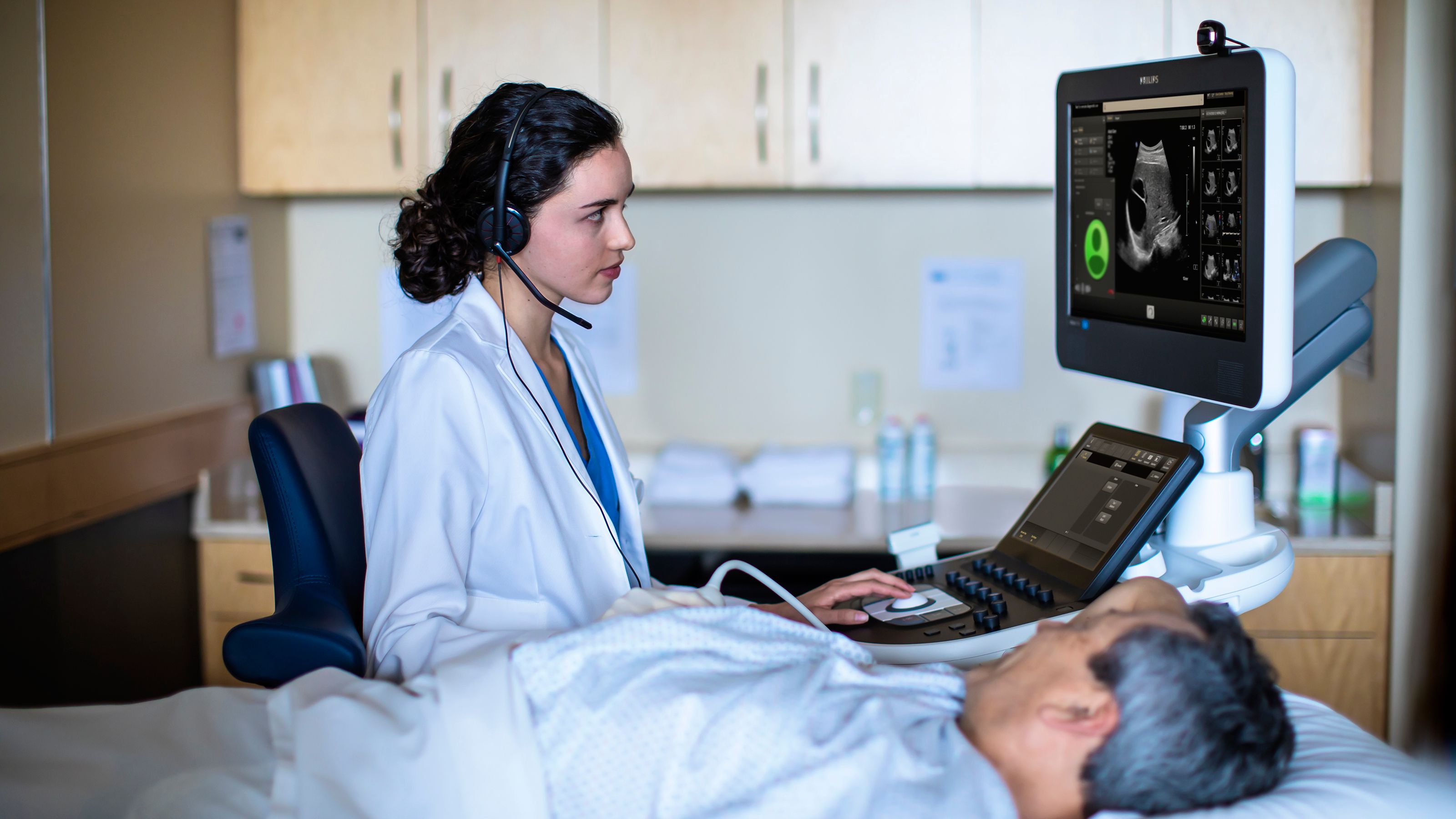
Philips Collaboration Live* enables secure talk, text, screen share and video stream functions, allowing users to remotely access expertise directly from the ultrasound system. The Collaboration Live solution can be used to provide real-time remote clinical diagnosis,** decision support and training on care protocols.

High-resolution transducers for swift and precise imaging
Philips offers a wide range of transducers for excellence in echocardiography, doppler and sonography. They feature intelligent imaging technology for 2D, 3D, 4D, flat, static and moving imaging along with an ergonomic design for comfortable scanning. For example, leading-edge xMatrix technology enables quick and easy volume acquisition, supports multiple interrogation capabilities, and provides views not possible with 2D imaging.

It’s compact without compromise
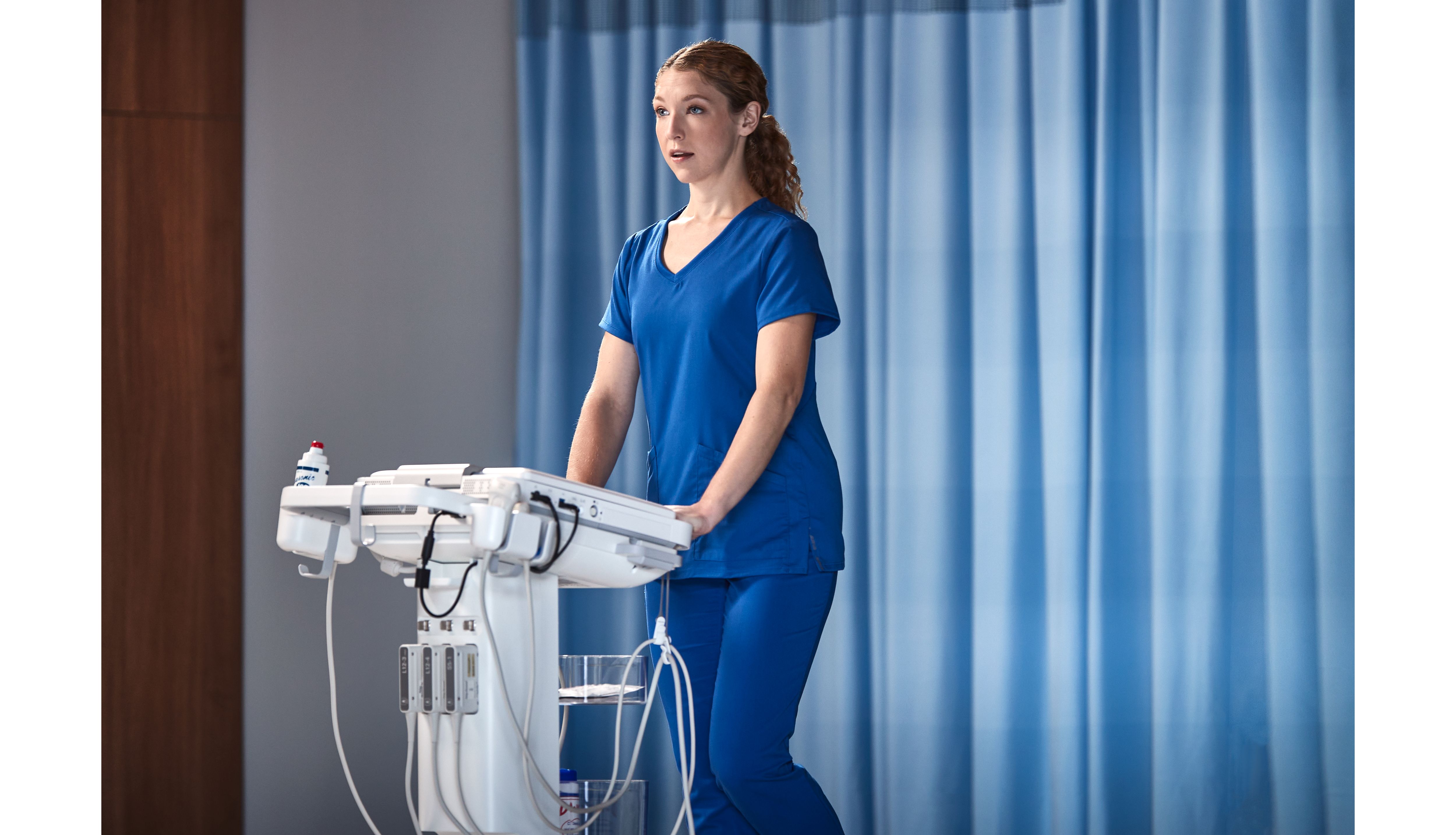
Compact Ultrasound System 5000 series brings full functionality and supports quick, confident answers to you, wherever you are. The Compact 5000 Series offers a feature-rich core and a versatile range of diagnostic solutions – all built into a highly portable, cleanable, easy-to-use system.

Enabling technologies for Ultrasound
Disclaimer
*Collaboration Live requires VM 7.0.5 or higher. Remote diagnostic use requires VM 9.0 or higher.
**Accessible via compatible clients including iOS, Android, Chrome web browser and Windows with release 9.0 or higher.


