Ultrasound
General imaging ultrasound
One platform. One user experience.
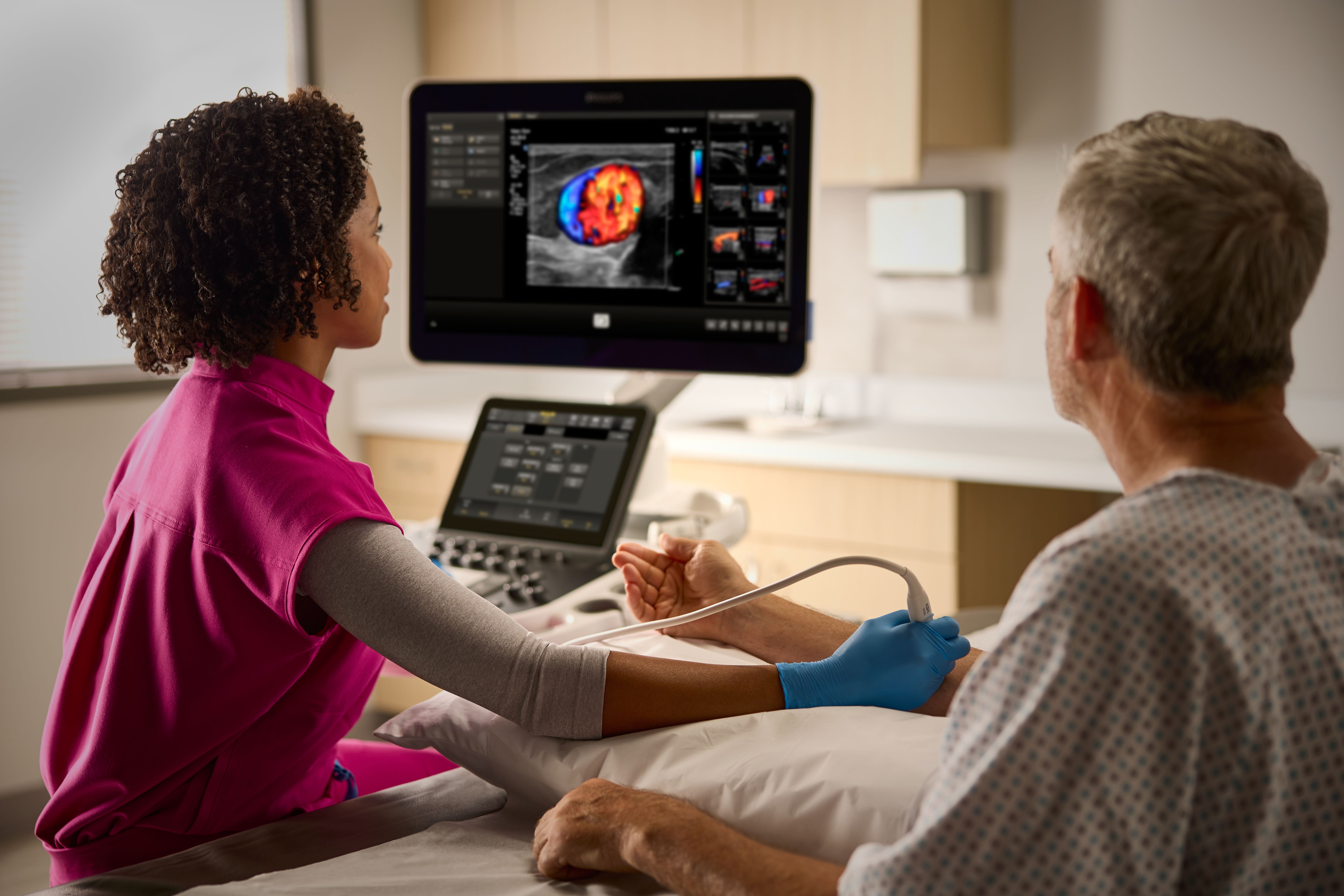
Designed to empower clinical decisions, enhance patient experiences and support efficiency, our portfolio of smart, connected General Imaging ultrasound systems deliver people-centric workflows, a shared platform and interface across our systems to streamline training and staffing flexibility, and image quality that sets us apart.

The power of one ultrasound
One product family with shared transducers, workflow and user interface, across a multitude of clinical applications.
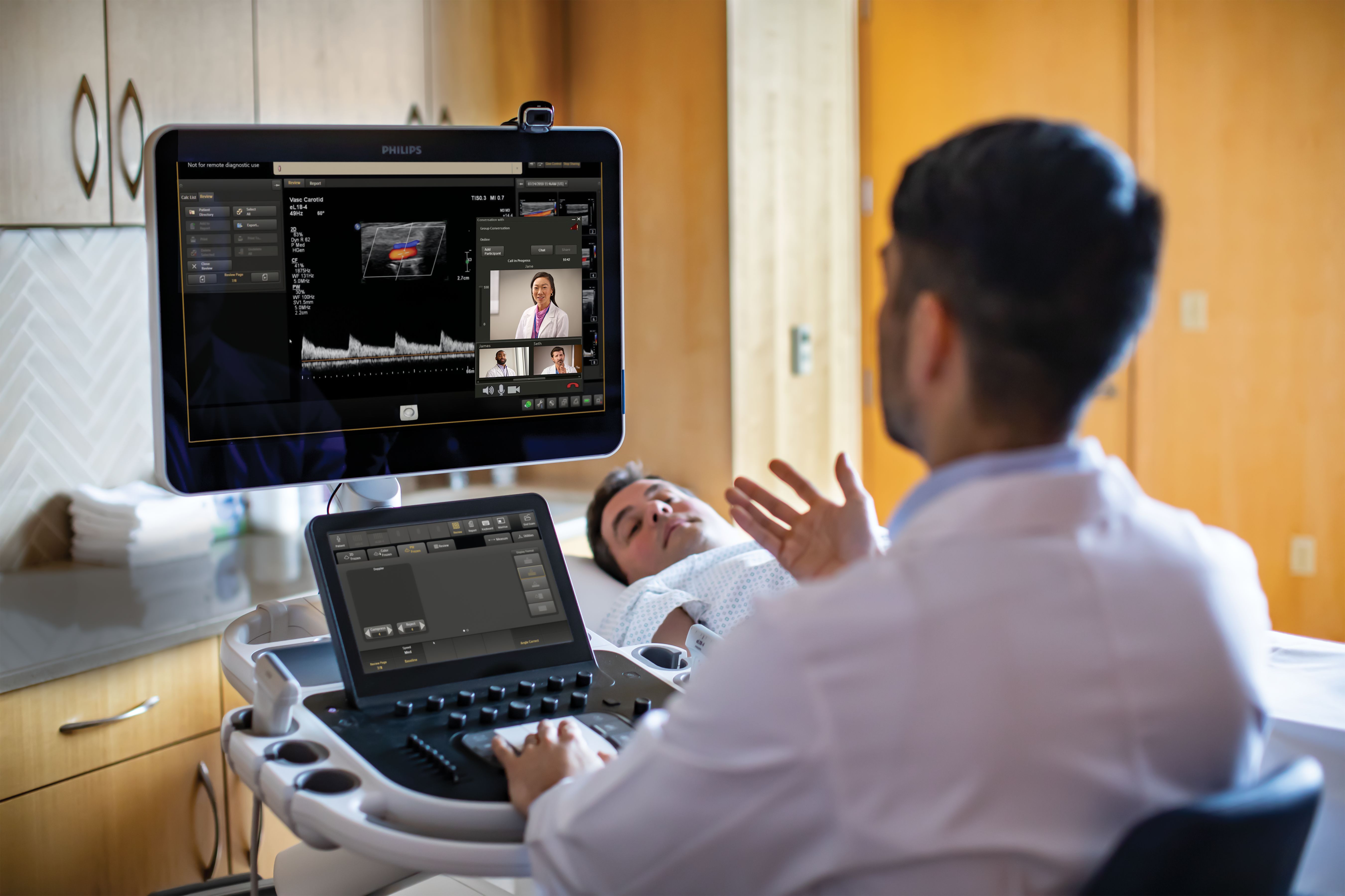
Collaboration Live
Extend your team without expanding it. Quickly and securely talk, text, screen share and video stream directly from an ultrasound system to a PC or mobile device, giving up to six care team members the ability to collaborate.

High-resolution transducers for swift and precise imaging
Philips offers a wide range of transducers for excellence in doppler and sonography. They feature intelligent imaging technology for 2D, 3D, 4D, flat, static and moving imaging along with an ergonomic design for comfortable scanning. For example, the award-winning mL 26-8 ultra-high frequency, compact linear array transducer allows you to image from eyeball to hip - all with the same transducer.

Technology Maximizer
With Technology Maximizer Plus, you automatically receive updated innovations for your systems as the updates are released, keeping your systems at the leading edge of clinical and operational value.

Enabling technologies for General imaging ultrasound
Related procedures
Disclaimer
Results are specific to the institution where they were obtained and may not reflect the results achievable at other institutions. Results in other cases may vary.


