Left atrial appendage occlusion
Image-guided therapy

Left atrial appendage occlusion

Solutions for left atrial appendage occlusion (LAAO)

Image-guided therapy solutions enhance confidence in left atrial appendage occlusion procedures (LAAO) with real-time visualization, efficient workflow set-up and streamlined decision-making.

Demonstrated results for left atrial appendage occlusion

  1. 100%

    physician agreement

    100%

    physician agreement

    physicians who participated in a simulated use study agreed that FlexArm offered uncompromised access to the patient’s head end suite1.

    Download
  2. 91%

    less transseptal puncture time

    91%

    less transseptal puncture time

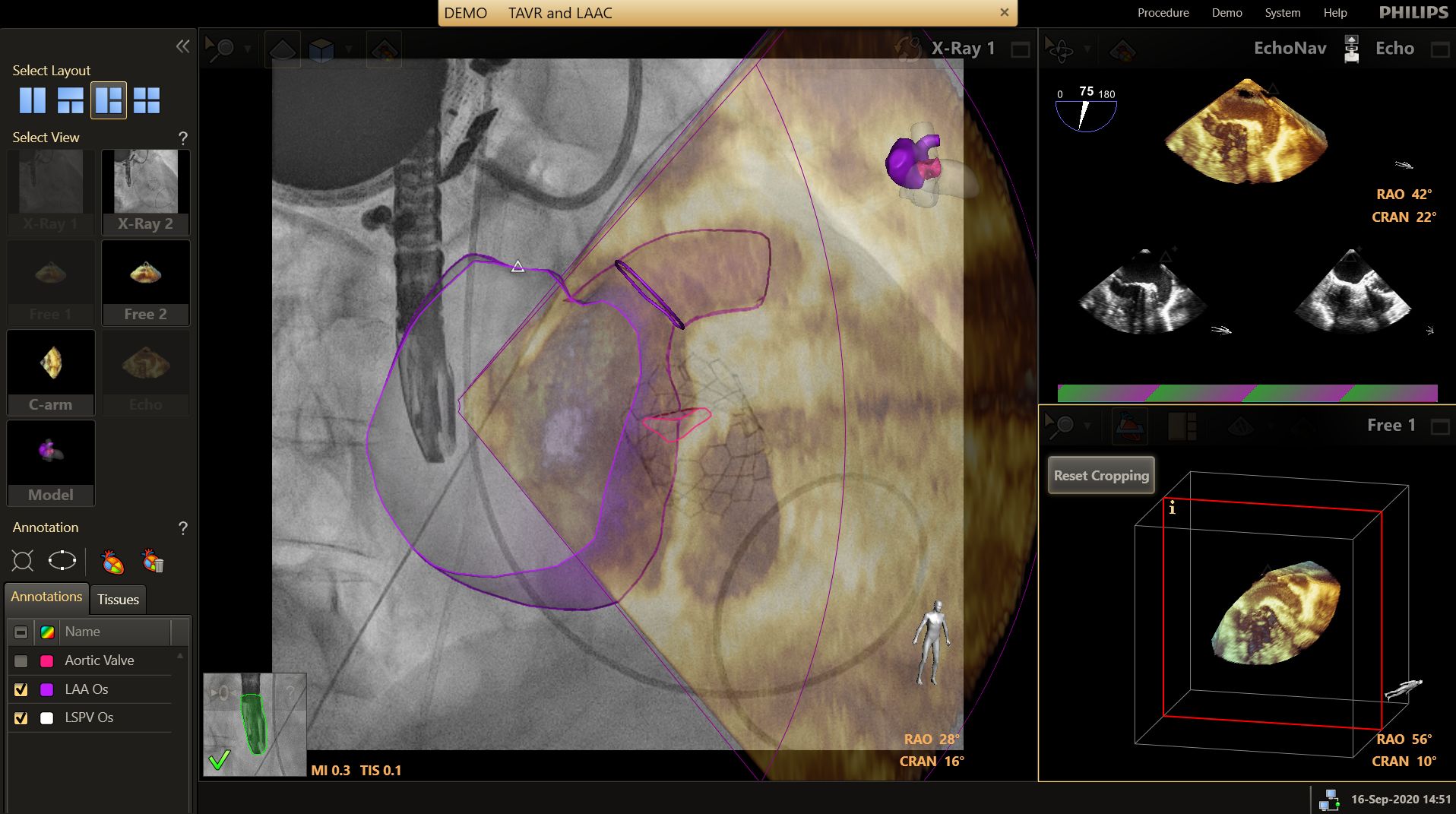
    Echonavigator reduces transseptal procedure time2

    Download
  3. 52%

    reduction in radiation dose

    52%

    reduction in radiation dose

    EchoNavigator reduces radiation dose by 52% in LAAO3.

    Read more
  4. 94%

    of clinicians say EPIQ MultiVue could help reduce risk of incorrect sized device

    94%

    of clinicians say EPIQ MultiVue could help reduce risk of incorrect sized device

    of clinicians who saw the new EPIQ CVxi thought the EPIQ MultiVue real-time alignment solution could help to reduce the risk of choosing an incorrectly sized device during interventional procedures.4

    Read more

Increase confidence

Philips offers integrated solutions for the treatment of LAAO, combining interventional X-ray, 3D ultrasound and interventional tools designed to enable confident navigation and positioning of devices to optimize outcomes.

Image-guided therapy can help simplify LAAO workflows

  • Gain optimal patient access during the procedures.
  • Achieve superb image quality at significantly lower dose across clinical areas, patients and operators.
  • Simplify pre-op structural heart disease planning and enhance insights with live fusion.
  • Enhance confidence in device manipulation and in understanding anatomical structures with EchoNavigator. 
  • Improve efficiency through the automation of imaging views and fusion.
  • Continuously monitor patients and improve Cath lab workflows with hemodynamic monitoring and data management.

LAAO procedure planning capabilities and imaging guidance

  • Flexible gantry positions optimize workflow with FlexArm on Azurion. 
  • Achieve high-quality imaging with excellent visibility at a low X-ray dose with ClarityIQ imaging technology.  
  • Reorient and combine ultrasound and X-ray information into a single augmented visualization for guidance and device-tissue relationship assessment with EchoNavigator. 
  • HeartNavigator offers fully automated CT analysis, including sizing and optimal X-ray projection, as well as Cath lab integration for automatic views and fluoroscopic fusion of planning information.

Related procedures

Footnotes
  1. Compared to the suites with Azurion 7 M20. Evaluated with clinical users in a simulated lab environment after approximately 20 minutes of practicing C-arm and table positioning.
  2. Afzal S, Veulemans V, Balzer J, Rassaf T, Hellhammer K, Polzin A, Kelm M, Zeus T. Safety and efficacy of transseptal puncture guided by real-time fusion of echocardiography and fluoroscopy. Neth Heart J. 2017 Feb;25(2):131-136. doi: 10.1007/s12471-016-0937-0. PMID: 27966185; PMCID: PMC5260626.
  3. Jungen et al., PLoS One 2015 Oct 14;10(10); Left Atrial Appendage Closure Guided by Integrated Echocardiography and Fluoroscopy Imaging Reduces Radiation Exposure
  4. Based on responses from 38 respondents
  5. Results from study conducted at St. Antonius Hospital. Results verified by NAMSA, independent third-party expert on study design and analytics. Results are specific to the institution where they were obtained and may not reflect the results achievable at other institutions.
Disclaimer
Please note that not all products displayed on our website may be available in your country due to varying local regulations and market conditions. For the most accurate availability information, please contact your local representative.