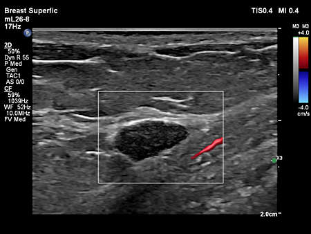
EPIQ Elite
Performance redefined
  • Philips EPIQ Elite ultrasound
  • Product in Use Photograph
  • Philps EPIQ Elite premium ultrasound
  • Product in Use Photograph
  • Product in Use Photograph
  • Product in Use Photograph
  • Standard product photograph

EPIQ Elite

Performance redefined
EPIQ Elite Elevate provides high-quality imaging and tailored clinical information to help clinicians deliver timely, confident answers to more patients worldwide. With advanced intelligence and an exceptional level of performance, EPIQ Elite meets the demands of today’s most challenging practices.

Clinical image gallery

  • eL18-4 scanning on a breast lesion.
    eL18-4 scanning on a breast lesion.
  • Affiniti VM10.0 C5-1 Abd Gen Liver Fatty LFQ
    Affiniti VM10.0 C5-1 Abd Gen Liver Fatty LFQ
  • Fetal Heart CPA with Flow Viewer
    Fetal Heart CPA with Flow Viewer
  • mL26-8 paired with Flow Viewer, scanning on a thyroid
    mL26-8 paired with Flow Viewer, scanning on a thyroid
  • L12-3 paired with CEUS Auto Scan, scanning on the Vascular Carotid Preset.
    L12-3 paired with CEUS Auto Scan, scanning on the Vascular Carotid Preset.
  • Ovary MFI Flow Viewer
    Ovary MFI Flow Viewer
  • Affiniti VM10.0 L12-5 Adv Breast 2D
    Affiniti VM10.0 L12-5 Adv Breast 2D
  • mL26-8 paired with Flow Viewer, scanning on a breast lesion
    mL26-8 paired with Flow Viewer, scanning on a breast lesion
  • Uterus Color Flow with Flow Viewer
    Uterus Color Flow with Flow Viewer
  • Ovarian blood flow with MFI using C10-3v transducer
    Ovarian blood flow with MFI using C10-3v transducer
  • Placenta CPA with Flow Viewer
    Placenta CPA with Flow Viewer
  • eL18-4 paired with CEUS, scanning on a thyroid.
    eL18-4 paired with CEUS, scanning on a thyroid.
  • Color Flow with Flow Viewer using C10-3v showing ovary perfusion
    Color Flow with Flow Viewer using C10-3v showing ovary perfusion
  • Flow Viewer applied to MFI with C10-3v uterus
    Flow Viewer applied to MFI with C10-3v uterus
  • L12-3 Ergo scanning on a calf vein
    L12-3 Ergo scanning on a calf vein
  • Umbilical cord shown with Color Flow with Flow Viewer using C5-1 transducer
    Umbilical cord shown with Color Flow with Flow Viewer using C5-1 transducer
  • C5-1 paired with Flow Viewer, scanning on the Pediatric Abdomen
    C5-1 paired with Flow Viewer, scanning on the Pediatric Abdomen
  • Fetal Aortic Arch CPA with Flow Viewer
    Fetal Aortic Arch CPA with Flow Viewer
  • Strain elastography demonstrating increased stiffness of an intrauterine fibroadenoma
    Strain elastography demonstrating increased stiffness of an intrauterine fibroadenoma
  • Umbilical Cord Color Flow with Flow Viewer
    Umbilical Cord Color Flow with Flow Viewer
  • Aortic arch CPA with Flow Viewer using C9-2 transducer
    Aortic arch CPA with Flow Viewer using C9-2 transducer
  • Affiniti VM10.0 L12-5 Adv Breast 2D
    Affiniti VM10.0 L12-5 Adv Breast 2D
  • Pulmonary vein and fetal heart MFI with Flow Viewer using C5-1 transducer
    Pulmonary vein and fetal heart MFI with Flow Viewer using C5-1 transducer
  • Fetal Lung Perfusion Color Flow with Flow Viewer
    Fetal Lung Perfusion Color Flow with Flow Viewer
  • Ovary Color Flow with Flow Viewer
    Ovary Color Flow with Flow Viewer
  • Affiniti VM10.0 L12-5 MSK Gen Biceps Tendon
    Affiniti VM10.0 L12-5 MSK Gen Biceps Tendon
  • C9-2 scanning on the Abdomen Renal Preset
    C9-2 scanning on the Abdomen Renal Preset
  • Auto Scan Of
    Auto Scan Of
  • Umbilical Cord Color Flow with Flow Viewer
    Umbilical Cord Color Flow with Flow Viewer
  • MSK Superfic Bowel FV
    MSK Superfic Bowel FV
  • C9-2 scanning on a liver.
    C9-2 scanning on a liver.
  • L12-3 Ergo paired with Flow Viewer, scanning on a carotid
    L12-3 Ergo paired with Flow Viewer, scanning on a carotid
  • EPIQ Elite Clinical Image
    EPIQ Elite Clinical Image
  • Fetal heart four-chamber color flow with Flow Viewer
    Fetal heart four-chamber color flow with Flow Viewer
Features
Service
The need to do more with less, rising case complexity and additional care settings put challenges with staffing, skill variability and standardization into sharp focus. This is where our services and solutions can help - get service tailored for your needs with our RightFit contracts, clinical and technical education and training to keep skills fresh, maximize your equipment investment with Technology Maximizer, and extend your team without expanding it with Collaboration Live.
Service
Auto Doppler
Automatically adjusts for optimal flow sensitivity and resolution, reducing 10 steps to 3 steps and also reducing the number of repetitive button pushes by an average of 68%.
Auto Doppler
MFI HD
Designed to detect slow and weak blood flow anatomy in tissue. This proprietary approach overcomes many of the barriers associated with conventional methods to detect small vessel blood flow with high resolution and minimal artifacts. MicroFlow Imaging maintains high frame rate and 2D image quality while applying advanced artifact reduction techniques to reveal small vessel anatomy
MFI HD


Specifications
  • Common Specifications
    Width
    60.6 cm/ 23.9 in
    Height
    146-171.5 cm/ 57.5-67.5 in
    Depth
    109.2 cm/ 43 in
    Weight
    104.3 kg/ 230 lb without peripheral devices
  • Control panel
    Monitor size
    24 inch / 60.96 cm HD display

Documentation

EPIQ Elite Elevate brochure
PDF|14.24 MB
EPIQ Elite & Affiniti ultrasound Interventional Radiology flyer
PDF|3.64 MB
EPIQ Elite & Affiniti Pediatric flyer
PDF|5.02 MB
EPIQ Elite & Affiniti ultrasound Vascular flyer
PDF|4.31 MB


Related products

  • mL26-8 Transducer Compact and Ultra-high frequency Transducer
    Discover the award-winning Philips mL26-8 ultra-high frequency compact linear array transducer, designed to provide exceptional imaging versatility from head to hip. With specialized presets for MSK, breast, vascular, dermal, and ocular applications, the mL26-8 offers unmatched adaptability on EPIQ & Affiniti. Proud recipient of the 'Best Innovation Award in General Imaging' at Journées Francophones de Radiologie 2023.
Disclaimer
*based on a sample size of 20 users
***Compared to previous capability