MRCAT Pelvis
MR-RT clinical application

MRCAT Pelvis
MR-RT clinical application
MRCAT Pelvis lets you plan radiation therapy using MRI as a single modality solution. Within just one MR exam, MRCAT Pelvis provides excellent soft-tissue contrast for target and OAR delineation, and continuous Hounsfield units for dose calculations.
MRCAT (MR for Calculating ATtenuation) data can be used for export to treatment planning systems for CT-equivalent** dose calculations. In addition, MR-based imaging enables CBCT-based positioning based on soft-tissue contrast with the look and feel of CT.
Clinical image gallery
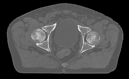
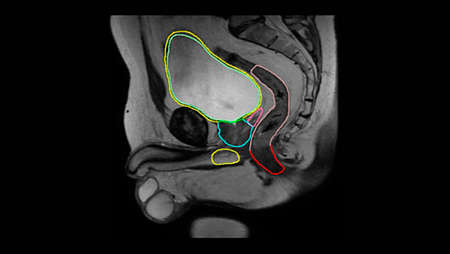
- MRCAT vs CT image
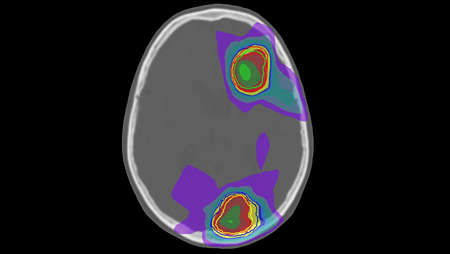
- MRCAT dose distribution
- Large field of view imaging
- MRCAT Pelvis with Continuous Hounsfield units
- MRCAT-based dose plan


Features
Unleash the real power of MR simulation
MRCAT Pelvis lets you plan radiation therapy for male and female pelvic cancer patients with soft-tissue tumors using MRI as a single-modality solution. This not only extends the benefits of MRI’s outstanding soft-tissue contrast to radiotherapy planning, but it also eliminates arduous, error-prone CT-MRI registration from the process, reducing uncertainties and complexity.

Fast, consistent imaging protocol
The dedicated MRCAT Pelvis imaging protocol includes a single, high-resolution, multi-contrast mDIXON sequence as the source for MRCAT generation. This scan is accelerated by Compressed SENSE, promoting patient comfort by minimizing time in the scanner. Moreover, it is standardized to deliver consistent results. A complementary 3D T2W scan provides high geometric accuracy and high-resolution image quality to support accurate delineation of target and critical structures. The total imaging protocol takes less than 15 minutes.

Automatic generation of synthetic CT images
MRCAT images are automatically generated using the mDIXON scan as source. Embedded image post-processing runs in the background, parallel to image acquisition, adding no time to the scanning session. Smart, validated algorithms enable automatic tissue segmentation and assignment of continuous Hounsfield units to deliver MRCAT images with CT-like density information for dose calculations.

Specifications
- MRCAT Pelvis
- Compatibility MR system
- Ingenia 1.5T and 3.0T MR-RT, Ambition 1.5T MR-RT and Elition 3.0T MR-RT
Related products
- The state-of-the-art Ingenia MR-RT platform featuring Ambition 1.5T and Elition 3.0T MR systems meets specific RT needs by providing high-quality MR images acquired in the treatment position. Smoothly integrate MRI through a comprehensive solution that considers your whole workflow, even for MR-only radiotherapy.
- MRCAT Brain clinical application allows the use of MRI as the primary imaging modality for radiotherapy planning of primary and metastatic tumors in the brain without the need for CT. Detailed anatomical information for contouring and attenuation maps for dose calculations are both obtained from a single, submillimeter resolution 3D T1W mDIXON MR sequence. Artificial Intelligence (AI) is used for fast computation of continuous Hounsfield units directly on the MR console.
- As a plug-in clinical application to Ingenia MR-RT, MRCAT Prostate + Auto-Contouring provides attenuation maps and automated, MR-based contours of prostate and organs at risk in as little as 20 minutes – all in a repeatable ‘one-click’ workflow.

Disclaimer
*Accurate means: MRCAT image acquisition provides <lt/> ± 1 mm geometric accuracy of image data in <lt/> 20 cm Diameter Spherical Volume (DSV) and <lt/> ± 2 mm geometric accuracy of image data in <lt/> 40 cm Diameter Spherical Volume (DSV)*. * Limited to 32 cm in z-direction in more than 95% of the points within the volume
**The simulated dose based on MRCAT images does not differ (Gamma analysis criterion 3%/3mm realized in 99% of voxels within the PTV or exceeding 75% of the maximum dose) in 95% of the pelvic cancer patients when compared with CT-based plan for EBRT.