Detector-based spectral CT changes cardiac imaging at Hospital Nuestra Señora del Rosario
- Featuring
- Featuring
- August 28 2024
- Duration 4:26
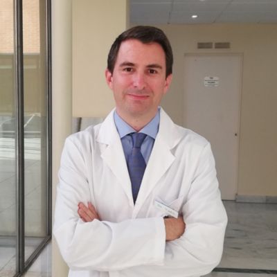
Dr. Eliseo Vañó Galván of Hospital Nuestra Señora del Rosario discusses what it’s like to use spectral-detector CT to increase diagnostic confidence, ease workflow, add value and advance patient care in cardiology. He believes the indication for spectral-detector CT will grow exponentially in the coming years.

At-a-glance:
- Spectral-detector CT is changing cardiac CT qualitatively
- Philips detector-based spectral CT more accurately characterizes atheromatous plaque, opening the door to CT myocardial perfusion imaging
- The system helps radiologists and cardiologists to work as a team, so they can focus on the patient
- The shift from multi detector CT to multi-energy CT offers a bright future for radiology
Dr. Eliseo Vañó Galván offers his perspective on spectral-detector CT
We know that the indication for cardiac CT is growing exponentially. I think this is a true change, a qualitative change that we have in the field of radiology and cardiac CT imaging. The advantage that the Philips detector-based spectral system has given us is that the spectral image is generated at the detector level, not at the emission level.
New opportunities in coronary imaging
This change means we don't have to choose when we make a spectral acquisition. Being at the detector level, the acquisition is always spectral, it is always on. We can reduce radiation doses in some cases because there is no need to perform scans without contrast. We don't have to repeat scans.
Now, as soon as we do coronary CT scans, we will be able to more accurately characterize the atheromatous plaque. This will open the door to something that we are not very accustomed to, which CT myocardial perfusion imaging. It really seems to me that Philips has made a change and focused on this type of spectral detection. It’s a qualitative change toward multi-energy imaging.
Patient-centric workflow
The Philips spectral CT is a system that is easy to implement in the daily workflow of a radiology department, so it allows us to really focus on what’s important, which is the patient and their diagnosis. It allows both radiologists and cardiologists to work as a team and for the patient to be the one who really benefits.
Leveraging new data
Another characteristic that will open many doors to us in the future is spectral information, which will provide a lot of data to do radiomic analysis. Radiomic consists of extracting image characteristics that the eye does not see, carrying out analyses and drawing conclusions regarding certain clinical data, prognoses of response to treatment, survival, etc.
Multi-energy CT is the future
Philips spectral CT offers possibilities that we cannot even begin to imagine. It’s going to open the door in cardiovascular pathology to defining prognostic aspects, treatment aspects, patient types, how to manage certain patients with cardiovascular disease, with coronary obstructions, with myocardial perfusion.
It will open the door to a better future for radiology and that’s why this qualitative change in shifting from multi detector CT to multi-energy CT is so important.

Read more
Dr. Eliseo Vañó Galván, President of the Spanish Society of Cardiothoracic Imaging, is Medical Director of CT/MRI at Hospital del Rosario, Madrid. A Stanford fellow, he leads research in 3T myocardial imaging and radiation dose optimization in cardiac CT.