iRotate electronic rotation and xPlane adjustable biplane imaging
Simultaneous multiplane imaging (SMPI) is a quick, simple to use and easy to understand echo modality that can help reduce scanning time in a high-volume echocardiography laboratory and provide unique clinical information on cardiac function and morphology. It also has the potential to ultimately bridge the gap between 2D and 3D thinking, helping to make the integration of 3D echocardiography easier in the clinical echo lab.
Although biplane and triplane echocardiography have been available since 1988, literature is currently scarce, suggesting the potential of this technique has not been fully appreciated, or the technology has not been adequate.
In 2003, transthoracic biplane echocardiography was introduced by Sugeng et al., showing the value of a simultaneous display of two imaging planes during stress echocardiography.2 This reduced examination time and had the potential for a single beat assessment of left ventricular (LV) function in patients with atrial fibrillation. The iRotate and xPlane modalities have the potential to overcome the limitations of biplane and triplane imaging and have important clinical value.
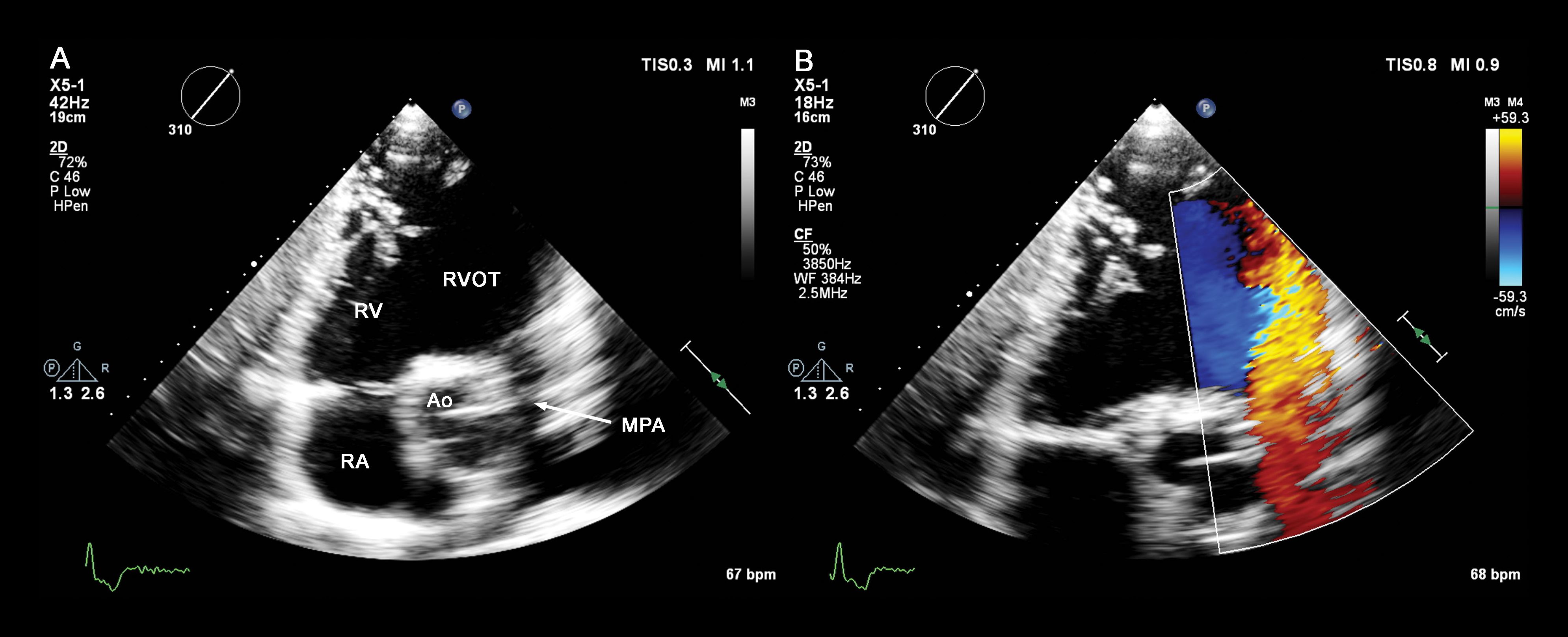
For the purposes of this paper, simultaneous multiplane imaging was performed using the Philips iE33 or EPIQ 7 ultrasound system, equipped with an X5-1 transthoracic echocardiography (TTE) or X7-2t transesophageal echocardiography (TEE) xMATRIX transducer. Rather than manually rotating the transducer to search for a non-obscured echo window, SMPI makes it possible to find the necessary view within the acoustical window without further manipulating the transducer.
The authors explain their imaging techniques, including helpful illustrations.
The paper details applications of SMPI, including automatic rotation during stress echocardiography, assessment of valve pathology, visualization of atrial septal defects and use of iRotate images for surgical and interventional cardiology.
