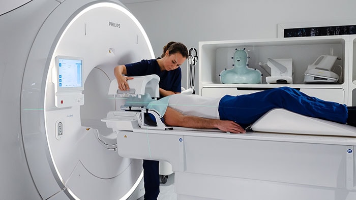
The Lyon University Hospital (Lyon, France) has recently been equipped with a Philips award-winning* Spectral CT 7500 scanner as part of a long-term strategic partnership agreement to provide the hospital with the latest state-of-the-art imaging solutions and facilitate joint research. Installed in the hospital’s imaging center, the use of this ground-breaking spectral CT imaging solution will help confirm Lyon University Hospital as a center of excellence in the management of cardiac and pulmonary disease, for which the Spectral CT 7500 is ideally suited.
“The long-term strategic partnership with Lyon University Hospital, which gives them access to the latest technological innovations in medical imaging, has already resulted in numerous scientific publications on the use of spectral CT,” said David Corcos, Precision Diagnosis Leader, Western Europe at Philips.
Since 2017, Lyon University Hospital has consistently been ahead of the curve in adopting spectral CT technology in clinical practice. Now equipped with the Spectral CT 7500 it will be able to leverage the benefits of Philips’ unique ‘always-on’ spectral CT imaging.
Faster diagnosis for more patients
The low radiation dose and reduced levels of contrast media injection achievable with the Spectral CT 7500 allow radiologists to manage a wide range of patients, including children, patients with renal failure who cannot therefore tolerate contrast agent, and patients with high body mass index (BMI). Overall, the solution contributes to increased access to care, faster and more accurate diagnoses, less need for second-line examinations, and smoother workflows. In studies, the use of Philips’ spectral CT technology resulted in a 26% reduction in the number of follow-up scans required because of an incomplete diagnosis [1], and a 34% reduction in overall time to diagnosis [2].
Unique ability
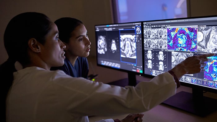
Unlike competitor offerings, the Spectral CT 7500’s dual-layer X-ray detector captures spectral information (imaging data at different X-ray energies) during a routine single scan, allowing radiologists to differentiate and quantify tissue with much greater precision by generating color-coded tissue type images. In an analysis by Seoul National University Hospital, Seoul, South Korea, its use resulted in a 23% increase in diagnostic confidence in terms of increased lesion conspicuity [3]. Because the scanner’s spectral data acquisition is always on, radiologists can view different tissue types without the need for re-scans. This unique ability is particularly relevant in the diagnosis of cardiovascular disease and neurovascular disorders for example, clearly locating the blood clot involved in a pulmonary embolism and areas of the brain affected by a stroke or heart attack. It is also useful in oncology and osteoarticular pathologies. The Spectral CT 7500 can perform most routine body scans in 2 seconds and routine head scans in less than 1 second.
It is now possible to carry out an exploration of the entire aorta in seconds, with minimal iodine injection.
Professor Philippe Douek
radiologist and project lead in Lyon University Hospital’s radiology department
"It is now possible to carry out an exploration of the entire aorta in seconds, with minimal iodine injection,” said Professor Philippe Douek, radiologist and project lead in Lyon University Hospital’s radiology department and guest speaker at Philips’ 3rd Annual Spectral CT and AI Virtual Summit (November 9, 2022).
Commenting on the convenience of the Spectral CT 7500’s always-on spectral data capture, radiographer Béatrice Rocher at the hospital’s imaging department said, “All the data is acquired in one go and remains available all the time. This is a real benefit because we can review the images in spectral mode at any time and further improve the quality of our diagnosis. In addition, the data acquisition system is unchanged compared to a standard acquisition, which is a real working comfort.”
Access to state-of-the-art equipment also promotes the attractiveness and retention of talent for our hospital and helps improve the quality of care in this region of France. One such example is the Spectral Photon CT Counting (SPCCT) project, an EU funded consortium of health systems and technology providers. During the project, Prof. Douek and team had access to the Philips Photon Counting prototype – a research system, not commercially available – which “…could benefit from the scientific advances of teams from various disciplines, but also that these researchers and practitioners benefit in return from the possibilities offered by this outstanding technology.” The SPCCT project focused on developing and validating a “…widely accessible, new quantitative and analytical in vivo imaging technology combining Spectral Photon Counting CT** and contrast agents, to accurately and early detect, characterize and monitor neurovascular and cardiovascular disease”. One outcome of the project was the first photon counting, human cardiac scan in the world.
Improving minimally-invasive treatment
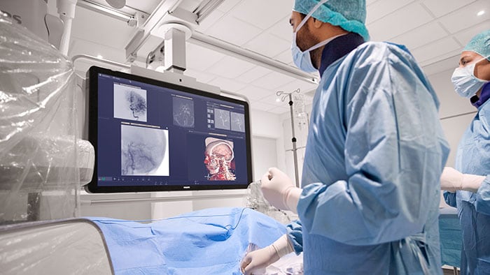
Diagnostic imaging is not the only area in which Philips’ Spectral CT 7500 is proving beneficial. By combining the Spectral CT 7500 system with the company’s Image Guided Therapy System – Azurion with FlexArm – Philips has developed a fully integrated hybrid angio CT suite solution for single-room, single-session diagnosis and treatment in areas such as oncology, stroke, and trauma care. Philips’ new Spectral Angio CT suite was showcased at the 2022 Cardiovascular and Interventional Radiological Society of Europe Annual Meeting (CIRSE 2022, September 10-14, Barcelona, Spain), where leading physicians shared the latest clinical insights on spectral CT imaging in interventional oncology during a Philips hosted symposium (registration required for on-demand viewing).
RSNA 2022
Philips will also spotlight its latest innovations in smart Connected Imaging Systems, including its Spectral CT technology, at this year’s upcoming Radiological Society of North America Annual Meeting (RSNA 2022, November 27 – December 1, Chicago, U.S.). Aimed at advancing clinical and diagnostic confidence, these innovations are designed to deliver clinically relevant, high-quality images that are right-first-time to enhance clinical decision-making and improve patient outcomes. Visit Philips on booth #6730 at RSNA 2022 or view Philips’ RSNA presence virtually via the Philips interactive online radiology experience. For other Philips updates during RSNA follow @PhilipsLiveFrom for #RSNA22 throughout the event.
[1] Analysis by LSU Health, New Orleans, LA, USA.
[2] Analysis by CARTI Cancer Center, Little Rock, AR, USA.
[3] Analysis by Seoul National University Hospital (SNUH), Seoul, South Korea
* Minnie Award for Best New Radiology Device
** Philips Photon Counting CT is for research only and is not a commercially available product
Share on social media
Topics
Contact

Kathy O'Reilly
Philips Global Press Office Tel.: +1 978-221-8919
You are about to visit a Philips global content page
Continue


