News center | United States


Just launched
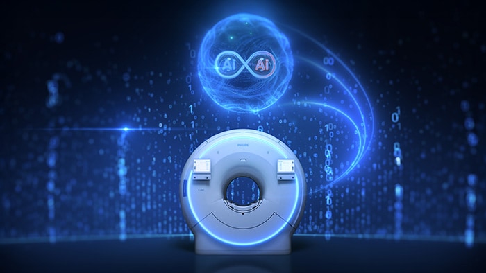
How can we unlock AI’s full potential in healthcare?
Learn new insights from patients and healthcare professionals in the 2025 Future Health Index - United States report.
Media updates: Philips Respironics voluntary June 2021 recall notification


Follow Philips news