xMATRIX Transducer Technology

xMATRIX Transducer Technology

is changing the exam experience

xMATRIX technology allows you to see more clearly, explore more fully and resolve more thoroughly, making exams faster and easier for both clinicians and patients.

*

Contact information

* This field is mandatory

*
*
*
*
*
*
*
*
*

By specifying your reason for contact we will be able to provide you with a better service.

We work with partners and distributors who may contact you about this Philips product on our behalf.

*
*

Final CEE consent

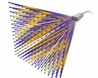
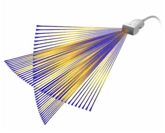
xMATRIX is our most leading-edge, versatile ultrasound transducer technology available today. It allows you to see more clearly, explore more fully and resolve more thoroughly, making exams faster and easier for both clinicians and patients. xMATRIX technology enables quick and easy volume acquisition, supports multiple interrogation capabilities, and provides views not possible with 2D imaging – and all with remarkable image quality. Confidently assess anatomy and function, easily identify abnormalities, and fully appreciate structural relationships in the 3D space.

xmatrix women health cogs

Enhanced workflow


With the touch of a button, xMATRIX offers all modes in a single transducer: 2D, 3D/4D, Live 3D Echo, Live xPlane, Live 3D Zoom, Live 3D Full Volume, iRotate, Live MPR, MPR, M-mode, Doppler, color Doppler, and CPA. No need to suspend acquisition to switch transducers. You can view ultrasound xMATRIX volume data on your PACs the same as you view CT and MR images. Once the data is acquired, the system captures the X, Y, Z, or iSlice MPR cineloops at the push of a button and sends them to your PACS. View, slice and interrogate at any time to suit your schedule.

architecture image

Extreme versatility


xMATRIX transducers support a wide range of clinical applications, including adult and pediatric cardiac exams; abdominal, vascular, obstetrical and gynecological exams; and cardiac and abdominal interventional procedures. xMATRIX is available on the EPIQ 7 and CX50 xMATRIX systems.

performance

Power and performance


xMATRIX transducers raise the bar of scanning performance. They harness the power of 150 computer boards, with fully-sampled elements ranging from 2,500 to over 9,000 for 360-degree focusing and steering. And when combined with Philips nSIGHT and PureWave crystal technologies, they are even more powerful.


Live volume imaging allows the acquisition and rendering of full volume data at true real-time frame rates with exceptional isovoxel resolution.

Live volume imaging
Live xPlane imaging


Live xPlane imaging creates two full-resolution planes simultaneously, allowing you to capture twice as much clinical information in the same amount of time as conventional 2D imaging.


Scanning comfort

While xMATRIX transducers are powerhouses of performance, they’re also easy to use. Their ergonomic design provides a slim, easy grip, for scanning comfort throughout the day.

Product image of Philips xMATRIX transducer

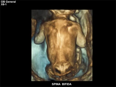
xMATRIX ultrasound: The fetal exam


The X6-1 xMATRIX ultrasound transducer supports all imaging modes for your fetal exams. Perform real-time 4D imaging of the fetal heart with image quality once reserved for 2D images. Implement elevation compounding with no frame rate penalty for enhanced speckle reduction and contrast resolution at all depths. Appreciate excellent 2D image clarity at shallow depths.

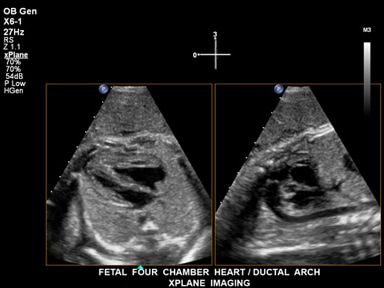
Quickly acquire fetal cardiac volumes with xMATRIX technology


iSTIC with xMATRIX allows you to acquire a high-resolution loop of volumes over a fetal cardiac cycle in as little as two seconds—versus 12 seconds with conventional STIC. Easily image very active fetuses to spot fetal heart anomalies early, reducing the need for costly retakes and saving your patients and staff the time and hassle of rescheduling exams.

spina bifida l new


The clarity of 3D fetal images may help you identify anomalies earlier and with more confidence, such as this case of spina bifida.


The additional information provided with Live xPlane imaging shows two views of the four-chamber fetal heart and ductal arch.

Fetal four chamber heart / ductal arch, xplane imaging
Experience with the X6-1 xMATRIX transducer

Experience with the X6-1 xMATRIX transducer


Jacques Abramowicz, Director of OB Ultrasound and Co-Director of Fetal and Neo-Natal Medicine Center, Rush University, Chicago, Illinois

You are about to visit a Philips global content page

Continue

You are about to visit a Philips global content page

Continue

Our site can best be viewed with the latest version of Microsoft Edge, Google Chrome or Firefox.